

Dinosaurs
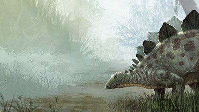
Dinosaurs were created by God on day six of creation, approximately 6,000 years ago. Dinosaurs were originally vegetarian. During the global flood, many were buried and fossilized, but two of each kind survived on Noah’s ark. Dinosaurs eventually died out due to human activity, climate changes, or other factors.
Contents
- When Did Dinosaurs Live?
- Types of Dinosaurs
- Dinosaurs and the Bible
- Behemoth in the Bible
- Leviathan in the Bible
- Humans & Dinosaurs Together?
- Soft Tissue Found in Dinosaurs
- Dinosaurs on Noah’s Ark?
- Dragons: Fact or Fable?
- Are Birds Evolved Dinosaurs?
- Dinosaur Movies
- What Happened to the Dinosaurs?
- Conclusion: Dinosaurs as “Missionary Lizards”
When Did Dinosaurs Live?
Evolutionists claim dinosaurs lived millions of years ago. But it is important to realize that when they dig up a dinosaur bone, it does not have a label attached showing its date. The Bible states that God made the land animals, including dinosaurs, on day six (Genesis 1:24–25), so they date from around 6,000 years ago.
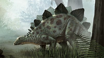
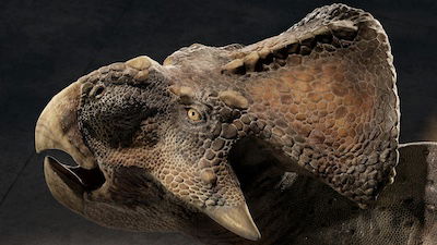
Types of Dinosaurs
Hypsilophodon foxii was one of the first dinosaurs discovered in 1849.
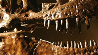
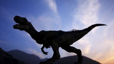
Tyrannosaurus rex, first described in 1905, this “tyrant lizard king” still holds the crown as the largest and most fearsome terrestrial predator ever discovered in North America.
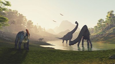
The sauropods (“lizard feet”) included the largest land animals in history.
A newly uncovered dinosaur, Albertaceratops nesmoi, had horns about three–feet long (located right above its eyebrows).

Dinosaurs and the Bible
As you add up all of the dates in the Bible, you’ll come to the conclusion that the creation of the earth and animals, including the dinosaurs, occurred only thousands of years ago, not millions of years ago. Thus, if the Bible is right (and it is!), dinosaurs must have lived within the past few thousands of years.
Behemoth in the Bible
Two great creatures, Behemoth and Leviathan, are described by God in Job. Some commentators have suggested behemoth was an elephant or hippo, but the description simply doesn’t match (e.g., behemoth “moves his “tail like a cedar”). It appears God is describing a sauropod dinosaur and a fearsome now-extinct sea creature.

Leviathan in the Bible
Though technically not a dinosaur but rather a marine reptile that may have also been able to come up onto land (Job 41:25 and 30), Leviathan can correctly be called a "dragon" which includes terrestrial, aquatic, and aerial reptiles.
In the biblical book of Job, God challenges Job by questioning his ability to capture Leviathan and make it his servant (Job 41:1–4). Leviathan is not a creature that little children can play with (Job 41:5) and is too large for traders to sell (Job 41:6).
God reminds Job that if a man is even thinking of capturing Leviathan with harpoons or fishing spears, then he needs to consider the battle that will take place (Job 41:7–8). If he does engage in battle with Leviathan, it will be the first and only time. Leviathan cannot be subdued by any man: this is a false hope, as he “is laid low even at the sight of him” (Job 41:9).

Humans & Dinosaurs Together?
Biblical creationists believe that man and dinosaurs lived at the same time because God said that he created man and land animals on day six. There is historical evidence of dinosaurs and man living together, such as the petroglyph in Natural Bridges, Utah, legends and stories of dragons in Europe, and the dragon motif in China. But one striking artifact in Asia is the bas-relief picture of a dinosaur in the ruins of Angkor outside of Siem Reap, Cambodia.
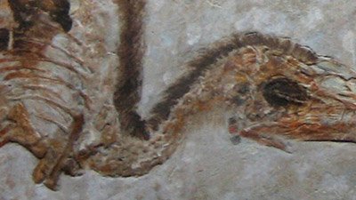
Soft Tissue Found in Dinosaurs
Dr. Mary Schweitzer and her team caught the world’s attention with a Science paper in 2005 that described intact blood vessels and red blood cells in a T. rex bone. But in fact, secular scientists have been reporting soft tissue in dinosaurs for decades in sometimes seldom-read technical literature.
Dinosaurs on Noah’s Ark?
In Genesis 6:19–20, the Bible says that two of every sort of land vertebrate (seven or seven pairs of the “clean” animals) were brought by God to the ark. Therefore, dinosaurs (land vertebrates) were represented on the ark.
Although there are about 668 names of dinosaurs, there are perhaps only 55 different “kinds” of dinosaurs. Furthermore, not all dinosaurs were huge like the Brachiosaurus, and even those dinosaurs on the ark were probably “teenagers” or young adults.

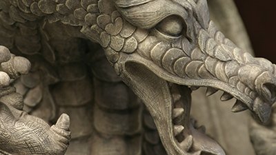
Dragons: Fact or Fable?
Ancient historians and writers clearly believed creatures like dragons were real. They describe seeing them first hand—often in the context of other types of animals that still live today. Some historians even describe the fiery flying serpents as real creatures in regions near where Moses and Isaiah were and point out the winged nature of these flying serpents. Such things are a great confirmation of the biblical text.
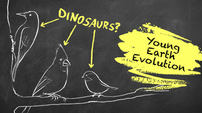
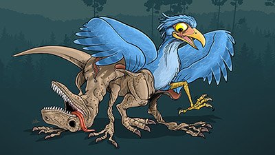
Are Birds Evolved Dinosaurs?
Having a true bird appear in the fossil record before alleged feathered dinosaurs, no mechanism to change scales into feathers, no mechanism to change a reptilian respiratory system into an avian respiratory system, and no legitimate dinosaurs found with feathers are all good indications that dinosaurs didn’t turn into birds. The evidence is consistent with what the Bible teaches about birds being unique and created after their kinds.
Genesis is clear that God didn’t make birds from pre-existing dinosaurs. In fact, dinosaurs (land animals made on day six) came after winged creatures made on day five, according to the Bible. Both biblically and scientifically, chicken-eaters around the world can rest easy—they aren’t eating mutant dinosaurs.
Dinosaur Movies
Television programs, thousands of books, and countless hours of research by thousands of qualified scientists have been devoted to studying dinosaurs. Is there an answer to their “mystery”? Sadly, many of the materials produced on the topic of dinosaurs teach parents and children evolutionary ideas and lead them away from believing the Bible.

What Happened to the Dinosaurs?
After Noah’s flood, around 4,300 years ago, the remnant of the land animals, including dinosaurs, came off the ark and lived in the present world, along with people. Because of sin, the judgments of the curse in Eden and the flood of Noah’s day have greatly changed earth over the past 6,000 years. Post-flood climatic change, lack of food, disease, and man’s activities caused many types of animals to become extinct. The dinosaurs, like many other creatures, died out.
Conclusion: Dinosaurs as “Missionary Lizards”
As Christians, we can use dinosaurs as “missionary lizards.” We can take what is popular with the culture and show how God’s Word explains it better. For example, soft tissue, like blood vessels and red blood cells, has been found in dinosaur bones. This soft tissue couldn’t last millions of years. The fossils confirm a young earth. We can use dinosaurs to help people trust the history of the Bible and also trust in the message of Jesus Christ that is also found in God’s Word.
Dinosaurs Topics
-
Dinosaur Bones
The presence of soft tissue in dinosaur bones cries out for a young earth, not for millions of years. These not-so-dry bones support the Bible’s history!
-

Dinosaur Extinction
An aura of mystery surrounds the extinction of the dinosaurs supposedly 65 million years ago—or does it? The Bible provides the true answer!
-
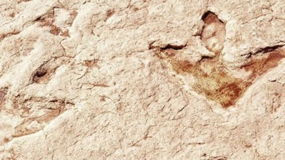
Dinosaur Footprints
Dinosaur bones aren’t the only artifacts left behind by these creatures. Fossilized footprints point toward the watery cataclysm that buried the dinosaurs.
-

Dinosaur Movies
Dinosaurs enthrall society, and many movies are produced to feed this fascination. But how much of these movies is fact and how much is evolutionary fiction?
-

Dinosaurs and Humans
Contrary to popular evolutionary opinion, the magnificent dinosaurs, created on the same day as Adam and Eve, recently lived and walked with humans.
-
Dragon Legends
Echoes of dinosaur and human cohabitation still resound around the world in dragon legends and myths.
-
Feathered Dinosaurs?
Did birds really evolve from dinosaurs? How should Christians understand the numerous claims of feathered dinosaurs used to support this idea?
-
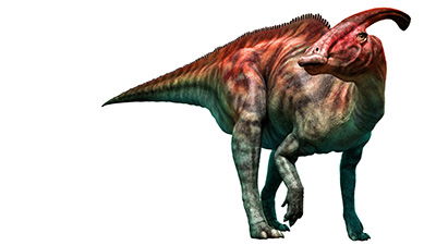
Types of Dinosaurs
Triceratops to T. rex, the many types of dinosaurs exhibit a great deal of variety. The design of these great reptiles calls out for a Creator.
-
When Did Dinosaurs Live?
Did dinosaurs live millions of years ago, or have these impressive creatures existed much more recently? Could some even be alive today?
Articles About Dinosaurs
-
June 11, 2026 from Ken Ham Blog
A recent headline declares, “Heron-Like, Fish-Eating Dinosaur from 70 Million Years Ago Discovered in Argentina.” What exactly does “heron-like” mean?
-
May 29, 2026 from Ken Ham Blog
It’s claimed that evolution explains everything, so what is the evolutionary explanation for how the T. rex, and other species like it, got those tiny arms?
-
May 11, 2026 from Ken Ham Blog
Did dinosaurs once swim across what’s now the Atlantic Ocean to colonize Africa?
-
Nov. 10, 2025 from Ken Ham Blog
One minute a 23-foot-long, 2,200-pound dinosaur was chomping on a crocodile leg, the next minute it was catastrophically buried.
-
Oct. 1, 2025 from Answers Magazine
Did dinosaurs and people even coexist? Could dinosaurs have fit in the ark?
-
Aug. 30, 2025 from Answers in Depth
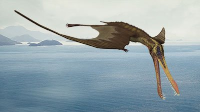
Depictions and documentation of dinosaurs, pterosaurs, and large marine reptiles living with man
-
July 28, 2025 from Ken Ham Blog
Genesis teaches that originally every living creature was vegetarian—eating the plants, seeds, and fruits that God created.
-
April 1, 2025 from Answers Magazine
Through imagination and technology, we are practicing the dominion mandate and may perhaps hear the calls of dinosaurs again.
-
Oct. 1, 2024 from Answers Magazine
At the Creation museum, Christian paleoartists are piecing together the past. How do they know if their presentation of extinct creatures matches created reality?
-
March 21, 2024 from Ken Ham Blog
A recent news article discussed research on how dinosaurs survived the end-Triassic/early-Jurassic mass extinction.
-
Dec. 18, 2023 from Ken Ham Blog
A recent discovery of the fossilized contents of a “teenage” tyrannosaur’s stomach gives researchers a glimpse into exactly what these massive reptiles ate.
-
Dec. 14, 2023 from Ken Ham Blog
According to a recent study, humans don’t live to 200 years old any longer because of dinosaurs.
-
Dec. 3, 2023 from Answers Magazine
Are dinosaurs proof of millions of years of evolution? Or a reminder of God’s glory?
-
Nov. 27, 2023 from Ken Ham Blog
The first line of a recent news article stated, “A new study argues that a real-life ‘Jurassic World’ could currently exist, just on another planet.”
-
April 29, 2023 from Answers in Depth
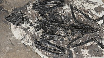
Cratonavis zhui is an alleged link between dinosaurs and birds.
-
April 13, 2023 from Ken Ham Blog
The image we all have in our minds of this top predator may be changing due to a new study that claims T. rex had lips!
-
Feb. 10, 2023 from Should Biblical Creation Become More Like Evolution?
Examining creationist arguments about the relationship between birds and dinosaurs
-
Sept. 29, 2022 from Ken Ham Blog
A new study claims they’ve identified the “earliest known mammal,” moving back the “appearance of mammals by about 20 million years.”
-
Aug. 21, 2022 from Answers Magazine
The evolutionary “family tree” that ties all dinosaurs together was recently redrawn. What bigger questions does this raise?
-
June 29, 2022 from Answers in Depth
This article is a warning to Christians to be discerning when it comes to accepting creationist research, just as we would be with other research.
-
June 2, 2022 from Ken Ham Blog
With an estimated 30-foot wingspan, the newly discovered “dragon of death” would’ve been a sight to behold—the largest pterosaur ever uncovered in South America.


Support the creation/gospel message by donating or getting involved!

Answers in Genesis is an apologetics ministry, dedicated to helping Christians defend their faith and proclaim the good news of Jesus Christ.
- Customer Service 800.778.3390
- Available Monday–Friday | 9 AM–5 PM ET
- © 2026 Answers in Genesis