
Are “Vestigial Organs” Really “Proof of Evolution . . . on Your Body”?
News to Know
“Proof of Evolution That You Can Find on Your Body,” a YouTube video reporting over 14 million hits in its first two weeks online, is the latest presentation to prop up the human evolutionary story through tales about vestigial structures, our presumably useless body parts.1 Ever since Darwin branded organs he considered nonfunctional as evidence for his beliefs, drumbeaters for the evolutionary worldview periodically trot out lists of vestigial organs to convince people that humans are the advanced animal products of random chance processes and natural selection.
This YouTube video—as you can see below—opens by declaring the human body a museum of natural history, loaded with “parts that aren’t there because you need them, but because your animal ancestors did” (:06), and claiming, “These remnants of our deep history only make sense within the framework of evolution by natural selection” (:16). The so-called vestigial organs, or “remnants,” selected for this video are structures and reflexes that are easily visible: a tendon in the forearm, seemingly useless muscles attached to your ears, goose bumps, the tailbone you may have bumped in a fall, and a tiny baby’s strong grasp. We contend that these and all other alleged anatomical footprints of our supposed evolutionary past are best understood as footprints of our wise Creator’s handiwork.
Palmaris Longus Muscle
Because it is one of several muscles that flex your wrist, the palmaris longus is a muscle you can do without. In fact, you may be among the 10–15% of people who already do without it in one or both arms. To see whether you have one or not, touch your pinkie to your thumb while flexing your wrist. If you have a palmaris longus, you will see its tendon as a tight, raised band extending down into your hand, where it attaches to the strong, flat layer of connective tissue in your palm.2
The palmaris longus, according to many reports, is quite unnecessary, its absence presumably causing no difference in grip strength. And being long, strong, and dispensable, this “vestigial” structure is a favorite among surgeons who harvest it for tendon grafts. But we must point out that the fact that a structure can be done without does not mean it is a footprint—a vestige—of an evolutionary past. Such a claim presumes we evolved from animals; it does not prove that we did.
The fact that a structure can be done without does not mean it is a footprint—a vestige—of an evolutionary past. Such a claim presumes we evolved from animals; it does not prove that we did.
Many of our body’s movements involve muscles that work together. The human wrist has more than one muscle available to flex it, and the palmaris longus’ contribution to wrist flexion is fairly minor. However, recent studies suggest this much maligned muscle may actually contribute more than it has been given credit for. A 2012 study published in the journal Physiotherapy reports that high-performance athletes in sports requiring a strong grip were substantially more likely to have a palmaris longus than athletes performing at a lower level. And sports requiring a sustained grip were statistically more likely to have participants who had a palmaris longus, whether at the amateur or professional level.3
Furthermore, anatomists typically focus on the wrist-flexing function of this muscle and ignore its contribution to the thumb’s movement in a different direction. The palmaris longus, both anatomically and functionally, makes a significant contribution to the thumb’s strength. While much of the palmaris longus tendon fans out into the palm, the lateral portion of the tendon attaches to the surface of the meaty thumb muscle (the abductor pollicis brevis) that forms the bulky thenar eminence. (You can see this as the broad muscular mound below your thumb.) This is the muscle that pulls the thumb outward and then toward your pinkie, to a position perpendicular to the palm. In fact, if you have a palmaris longus, you can watch its tendon tense at the wrist as you move your thumb this way. A 2010 study published in Clinical Anatomy compared the strength of this motion in the two hands of people who had only one palmaris longus. Regardless of which hand was dominant, strength was significantly greater in the hand with a palmaris longus.4
While it is quite reasonable that a person requiring reconstructive surgery is well-advised to sacrifice his or her palmaris longus to the greater need, these studies recommend that surgeons and anatomists should apprise patients of the potential impact its loss might have on their athletic performance and also recognize the muscle’s important contribution to the strength of thumb abduction.
Only human arrogance presumes that just because we haven’t yet discovered a body part’s function that it has none, and that it is therefore a useless evolutionary leftover. Likewise, just because a structure is absent in some people or highly variable does not mean some people are farther up the tree of life from an ape-like ancestor than others. Those of us who have spent much time in a medical school anatomy laboratory or as surgeons navigating the variable anatomy of the human body should realize such differences have no evolutionary significance, but are only variations within the human species. Their existence only screams “evolution” to those who already believe that Darwinian evolution explains the origin of all our parts.
Despite the headline position of the palmaris longus in this video about vestigial traits, this evolutionary argument should be relegated to the vestigial trash heap, not this muscle.
Outer Ear Muscles
What about the muscles attached to each outer ear, three of which are pictured as vestigial structures in the video? My dog can express emotion with its ears, flatten them against his head when he’s frightened, and re-orient them toward new and interesting sounds. The rabbits in my yard tweak their ears in his direction as they monitor his stealthy approach. Humans and apes cannot do this. However, as the video points out, the electrical activity in these muscles spikes in response to sudden noises, and it does so on the side from which the noise originates. The narrator says this shows that we have lost the ability to redirect our ears through evolution.
It is interesting that this vestigial video repeatedly uses the loss of a structure . . . or function . . . as evidence for the upward evolution of new structures and functions.
The ear muscles’ neural circuitry in our brain also leads to a reflexive shifting of our eyes to the side from which a sudden noise comes. This reflex is accompanied by a slight curling of the ear’s outer edge.5 These associated reflexive responses suggest the outer ear muscles may have a purpose not yet understood by scientists. Our outer ear is shaped and positioned to help us determine the direction from which sound is coming. And as part of the startle reflex—and a part that clearly responds directionally—the outer ears’ muscles may well serve to help us direct our attention to the source of a sound even though we do not re-direct our ears like a rabbit or a dog.
It is interesting that this vestigial video repeatedly uses the loss of a structure—like the 10–15% of people who are simply born without one or both of their palmaris longus muscles—or function—like the supposed loss of ear-orienting ability in humans as distant ancestors climbed the tree of life—as evidence for the upward evolution of new structures and functions. This sort of thinking is a bit backwards, and purely a product of the evolutionists’ tendency to assume evolution happened and to interpret what they see in a way to fit their story. However, whenever a structure or a function is, over time, lost within a particular kind of animal, or among humans, there has been either a lack of expression or a loss of information. Such a loss in no way demonstrates that an evolutionary gain of information once happened in the deep past.
Goose Bumps
Goose bumps are also implicated in the YouTube video as proof that our bodies are covered with useless evolutionary leftovers. Goose bumps form when tiny muscles attached to our hair follicles tug on them to make our hairs stand up a little taller, a typical response to a chill or even to a strong emotion. The associated tug on the attached skin makes those little bumps pop up. Because we don’t have fur or feathers that fluff up to keep us warm or to make us look big and fearsome, evolutionists claim goose bumps are visible vestigial structures, proof of our evolutionary past.
There is certainly nothing useless about the equipment that produces goose bumps, and goose bumps are not the proof of an evolutionary past.
But are goose bumps, the fine hair covering most of our skin, and the hair follicles and tiny muscles attached to them really useless to us? Not at all! Those muscles that tug on our hair follicles help the protective oil produced by follicles’ sebaceous glands to ooze out onto the skin. Their muscle action can also generate a bit of additional heat on a cold day, though we can be thankful that we do not have fur to impede our evaporating sweat from cooling us on a hot day. And inside every hair follicle is a supply of cells that can transform into the raw material for healing when needed. Without this supply of epithelial cells, even minor wounds would have to slowly heal from the edges inward. Our hair follicles are also attached to sensitive nerve endings, and when strong emotions prompt our fine hairs to stand up, they are more easily touched, increasing somewhat our sensitivity to the brush of danger. There is certainly nothing useless about the equipment that produces goose bumps, and goose bumps are not the proof of an evolutionary past.
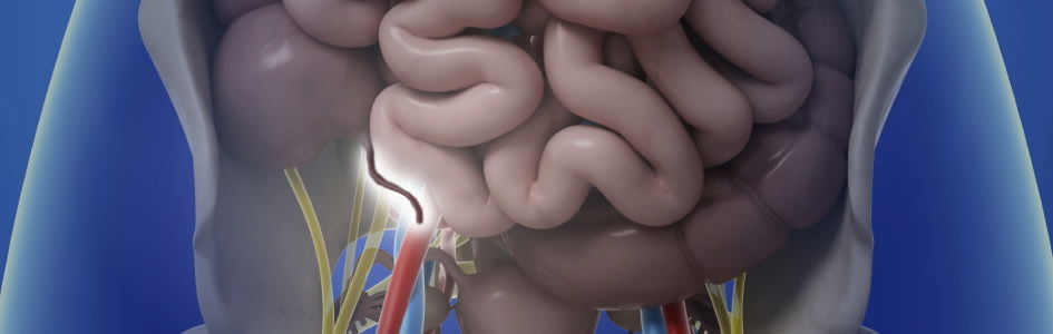
The Tail
What list of supposed footprints of our evolutionary past—a classic in the vestigial organ hall of fame—would be complete without a discussion of the human tailbone? This video does not disappoint, even including the overblown claim that “human embryos closely resemble embryos of other vertebrates” (2:58). (Do human embryos and other vertebrates have the same basic body plan? Yes. But the resemblance historically has been greatly—and even fraudulently—exaggerated. Learn more about it in “.”) The YouTube video’s narrator asserts that as the result of our “ancestral blueprint” (3:17), human embryos have “a tail with 10 to 12 developing vertebrae” (3:02) until programmed cell death destroys it, leaving the 3 to 5 vertebrae that comprise our coccyx.
Does the human coccyx at the end of the vertebral column hang us in our rightful place on the tree of life? Or is the coccyx a perfectly designed structure that serves our needs as well as a monkey’s tail serves its need to swing in the trees?
God designed humans to walk upright and gave the human coccyx a vital function to support our vertical lifestyle.
God designed humans to walk upright and gave the human coccyx a vital function to support our vertical lifestyle. The muscles of the pelvis attach to the coccyx to form slings supporting all the pelvic organs in their proper geometric orientation. When those muscles weaken, pelvic organs can herniate downward causing discomfort and a distressingly inconvenient loss of control. The coccyx, essential to our upright life, is not a useless evolutionary remnant.
Neither is the human embryo’s so-called tail evidence of an evolutionary past. From the third to the seventh week of development, the lower part of the curled, C-shaped body resembles a tail. It is not packed with 10 to 12 vertebrae as the video claims but with series of somites—blocks of cells that are designed to differentiate into muscles, bones, cartilage, and other supporting structures or to act as the scaffolding or stimuli for other structures to grow. Within this temporary so-called “tail” are a number of such somites, as well as a secondary neural tube and the lower part of the notochord. Cells in the notochord and secondary neural tube secrete molecular signals that guide somite differentiation. But that’s not all! The secondary neural tube that forms in this “tail” is specifically responsible for forming the fibrous support for all those spinal nerves that extend out from the end of the spinal cord to innervate the lowermost parts of the body. When development is complete, the parts no longer needed disintegrate and leave the fibrous support for the nerves in place. The development of the support structures for these nerves is certainly an important event in the life of an embryo! There is nothing useless about it. It is not a vestigial organ.
Palmar Grasp Reflex
Finally, the YouTube video winds up with what the narrator calls “the most adorable vestigial behavior”—the palmar grasp reflex (3:26). Until about six months of age, normal human babies tightly grip whatever presses on their palms. (A young baby’s feet also respond with a plantar grasp reflex by trying to curl around a finger pressing against the sole just behind the toes.) The video shows a film of a baby supporting its own weight, attributing its great grip to its “inner monkey.” (3:48)
Comparing this “vestigial trait” to the tight grasp of a primate animal baby on its mother, evolutionists have long claimed that our palmar grasp reflex is a vestige of our primate ancestral baby’s grip on its arboreal mother, a grip without which it would not only have lost its opportunity to suckle nourishment but also would have dropped to the ground as its mother moved through the trees. And because human mothers do not habitually swing through the trees with their clinging babies in tow, evolutionists consider the palmar (and plantar) grasp reflexes to be useless leftovers, evidence of our evolutionary past.
A study of the grasp reflexes in normal and abnormal babies reveals a different story, however. The palmar reflex occurs in all neurologically normal infants until about 3 to 6 months of age, thereafter decreasing in intensity and finally disappearing. The plantar reflex similarly disappears at 6 to 12 months of age. Adorable as the finger-clutching grip of a baby is to doting parents, grasp reflexes are the result of normal spinal reflexes that are in place in a baby long before birth.6
Later, as neuronal connections form in the growing baby’s nervous system, the brain begins to inhibit these particular spinal reflexes. Eventually, with increasing maturity of the nervous system, the grasping reflexes disappear altogether, being replaced by the ability to balance and to voluntarily use the hands and feet to manipulate objects and eventually to walk.
The reflexive grasp needs no “function” to justify its presence in human babies
Babies with neurological abnormalities may not lose their grasp reflexes at the time normal babies do.7 This persistence is pathological—a sign of an abnormality—but there is nothing primitive about it. Infant grasp reflexes merely reflect the normal spinal reflexes that are expressed in a maturing nervous system prior to the completion of its neuronal connections. The reflexive grasp needs no “function” to justify its presence in human babies, though the feeling of affection and dependence adults infer from having a finger tightly grasped may well serve a function in the psychological sense, facilitating the bond between parents and their helpless babies. Nevertheless, the determination of evolutionists to attribute this immature phase of normal neurological development to our supposed kinship with monkeys is a worldview-based just-so story with no scientific validity.
Vestigial Arguments
The word vestige comes from the Latin word for footprint. Vestigial organs—our supposed useless evolutionary leftovers—are popularly considered evolution’s footprints. But they are not. We humans think highly of our own intellect, but we are not all-knowing. How much baseless pride does it take to insist that a structure with an unknown function really has none, instead of just an undiscovered one? And a lost structure or function at most represents a loss of information, not sufficient evidence to prove that upward evolution of complexity ever occurred. Molecules-to-man evolution, including the many claims about the evolution of humans from an ape-like ancestor, are biblically inconsistent and scientifically insupportable.
These so-called vestigial structures and behaviors are not footprints of evolution.
These so-called vestigial structures and behaviors are not footprints of evolution, proofs of evolution visible on our own bodies. But they are footprints of a different sort. They are the footprints of the intricate development of the human embryo and of the finely engineered designs of our Designer, the Creator God of the Bible. Many of our unique human features would equip us poorly for life as animals, but they suit us well for our lives as human beings, enabling us to live the sort of lives God designed us for.
Anatomist, biologist, and creation scientist Dr. David Menton responds to popular YouTube video “Proof of Evolution That You Can Find on Your Body” (posted by VOX) from a biblical and scientific worldview.
Footnotes
- “Proof of Evolution That You Can Find on Your Body,” YouTube video, posted by “Vox,” March 17, 2016, https://www.youtube.com/watch?v=rFxu7NEoKC8.
- The palmaris longus muscle originates just above the elbow, from the upper arm bone. Tendons attach muscle to bones and other sturdy structures, and it is the muscle’s tendon that you will usually see if you have a palmaris longus. The muscle is highly variable even in people who have it. In some cases the muscular portion is the part seen at the wrist. And while many people lack this muscle, some people even have two.
- The 642 test subjects consisted of medical students and both amateur and professional athletes. The study focused on overall performance in sports that did or did not require strong or sustained grips. It did not attempt to break the grip down into its various muscular components but considered instead whether the palmaris longus made a statistically demonstrable contribution to overall performance. To learn more, see Craig Fowlie, Colin Fuller, and Margaret K. Pratten, “Assessment of the Presence/Absence of the Palmaris Longus Muscle in Different Sports, and Elite and Non-elite Sport Population,” Physiotherapy 98, no. 2 (2012): 138–142, doi:10.1016/j.physio.2011.02.006.
- Hope Gangata, Robert Ndou, and Graham Louw, “The Contribution of the Palmaris Longus Muscle to the Strength of Thumb Abduction,” Clinical Anatomy 23 (2010): 431–436, doi:10.1002/ca.20960.
- This 2–3 mm visible curling of the dorsal edge of the outer ear is a motion produced by a fourth outer ear muscle, the transversus auriculae. The three muscles pictured in the video—the auricularis anterior, the auricularis superior, and the auricularis posterior—are arrayed in front, above, and behind the outer ear. They exhibit electrical activity on the side from which a sudden sound comes at the same time as the ear’s edge curls and the eyes shift sharply in the appropriate direction. For more information see Steven Hackley, “Evidence for a Vestigial Pinna-Orienting System in Humans,” Psychophysiology 52, no. 10 (2015): 1263–1270, doi:10.1111/psyp.12501.
- The brain is not required, and even anencephalic babies exhibit the palmar and plantar grasping reflexes.
- Yasuyuki Futagi, Yasuhisa Toribe, and Yasushiro Suzuki, “The Grasp Reflex and Moro Reflex in Infants: Hierarchy of Primitive Reflex Responses,” International Journal of Pediatrics (2012): doi:10.1155/2012/191562.


Support the creation/gospel message by donating or getting involved!

Answers in Genesis is an apologetics ministry, dedicated to helping Christians defend their faith and proclaim the good news of Jesus Christ.
- Customer Service 800.778.3390
- Available Monday–Friday | 9 AM–5 PM ET
- © 2026 Answers in Genesis